Mapping the Fascicular Morphology and Organization of the Human Sciatic Nerve via High-Resolution MicroCT Imaging

Mapping the Fascicular Morphology and Organization of the Human Sciatic Nerve via High-Resolution MicroCT Imaging
Zhang, J.; Lam, V. H.; Nuzov, N. B.; Brunsman, B. A. S.; Pascol, T.; Onabiyi, A.; Prince, R.; Kalpatthi, H.; Gustafson, K.; Triolo, R.; Pelot, N. A.; Crofton, A.; Shoffstall, A. J.
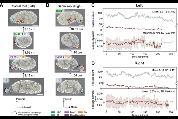
AbstractObjective. Implanted neuroprostheses can restore standing and walking after spinal cord injury and somatosensation after limb loss. Yet, current approaches often fail to sufficiently and reliably activate hamstring muscles crucial for robust upright mobility, or to target the afferent fibers in mixed nerves like the sciatic nerve for sensory neuroprostheses. We developed a novel methodology using high-resolution micro-computed tomography (microCT) imaging to visualize and track fascicle groups innervating distinct hamstring muscle targets along the human sciatic nerve. This approach offers an efficient tool for mapping fascicular topography in complex neural pathways, providing an anatomically informed framework for optimizing standing neuroprostheses. Methods. Bilateral sciatic nerves were dissected and excised from an embalmed human cadaver, annotated with branch names, and stained with phosphotungstic acid before undergoing microCT scanning at 11.4 m isotropic resolution. Resulting images were segmented with a 3D U-Net convolutional neural network. Segmentation results were used to quantify morphological metrics and track the fascicular organization along ~25 to 30 cm of the nerve. Results. Gross dissection revealed a proximal-to-distal branching sequence in the right and left sciatic nerves: long head of the biceps femoris (lhBF), hamstring part of the adductor magnus and semimembranosus (HAM/SM), and the semitendinosus (ST) muscles, with branches originating medially from the sciatic nerve and following an inferomedial trajectory. Branch-free lengths exhibited asymmetry, especially between the lumbosacral roots to the first branch (5.5 cm left vs. 1.5 cm right) and lhBF to the HAM/SM branch (9.0 cm left vs. 16.5 cm right). MicroCT analysis quantified bilateral symmetry in fascicle diameters (~0.4 mm) and total fascicle counts (~84). In contrast, hamstring-innervating fascicle counts were asymmetric, averaging fewer on the left (~7) than the right (~9). The 3D fascicular maps revealed that hamstring fascicles primarily aggregated within the anteromedial aspect of the sciatic nerve and remained separate for considerable distances (up to ~13 cm for ST) proximal to their branching points. Significance. Our microCT-based approach enables efficient, high-resolution 3D mapping of fascicular organization within large, complex peripheral nerves like the sciatic nerve, overcoming previous technical limitations. By quantifying fascicular morphology and organization, this methodology directly informs the development of neuroprostheses with enhanced activation of the hamstring muscle group, improving standing functions.