Female Anterior Cruciate Ligaments Exhibit a Muted Mechanobiological Response to Mechanical Loading

Female Anterior Cruciate Ligaments Exhibit a Muted Mechanobiological Response to Mechanical Loading
Paschall, L.; Konnaris, M.; Tabdanov, E. D.; Dhawan, A.; Szczesny, S.
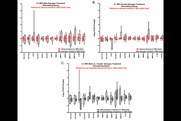
AbstractFemale athletes are significantly more likely to tear their anterior cruciate ligament (ACL) compared to their male counterparts. While there are several potential reasons for this, previous data from our lab demonstrated that female ACL explants have an impaired remodeling response to loading, which may prevent the repair of fatigue damage and lead to increased ACL rupture. The objective of this study was to identify the mechanisms driving the impaired remodeling of female ACLs to cyclic loading, including the role of estrogen. ACLs were harvested from male and female New Zealand white rabbits and cyclically loaded in a tensile bioreactor followed by bulk RNA-sequencing. Additional ACL explants treated with or without estradiol were analyzed using RT-qPCR to determine the regulatory effect of estrogen on markers for tissue remodeling and inflammatory cytokines with cyclic loading. We found that female ACLs exhibited significantly fewer differentially expressed genes (DEGs) in response to loading compared to male ACLs. Additionally, multiple mechanotransduction pathways were enriched with loading only in the male ACLs. While a few estrogen-related pathways were enriched in both male and female ACLs with loading, the expression of tissue remodeling markers was not different between estrogen treatment and vehicle control. Together, our findings highlight specific mechanotransduction pathways that may be responsible for the muted biological response of female ACLs to load, which provides a potential explanation for the increased rate of ACL tears in women.