3D imaging of human pancreas suggests islet size and endocrine composition influence their loss in type 1 diabetes
3D imaging of human pancreas suggests islet size and endocrine composition influence their loss in type 1 diabetes
Rippa, A.; Posgai, A. L.; Currlin, S.; Brusko, M.; Williams, M. D.; Kaddis, J. S.; Kusmartseva, I.; Wasserfall, C. H.; Campbell-Thompson, M.; Atkinson, M. A.
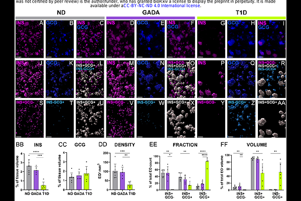
AbstractA high-definition description of pancreatic islets would prove beneficial for understanding the pathophysiology of type 1 diabetes (T1D), yet significant knowledge voids exist in terms of their size, endocrine cell composition, and number in both health and disease. Here, 3-dimensional (3D) analyses of pancreata from control persons without diabetes (ND) revealed heretofore underappreciated frequencies (approximately 50%) of insulin-positive (INS+) glucagon-negative (GCG-) islets. Non-diabetic individuals positive for a single Glutamic acid decarboxylase autoantibody (GADA+) yet at increased risk for disease consistently demonstrated endocrine features, including islet volume and cell composition, closely resembling the age-matched ND controls. In contrast, pancreata from individuals with short-duration T1D demonstrated significantly reduced islet density and a dramatic loss of INS+GCG- islets with preservation of large INS+GCG+ islets. The size and cellular composition of pancreatic islets may, therefore, represent influential factors that impact {beta}-cell loss during T1D disease progression.