Embryonic development of C. elegans sense organs
Embryonic development of C. elegans sense organs
Wexler, L.; Kolotuev, I.; Heiman, M. G.
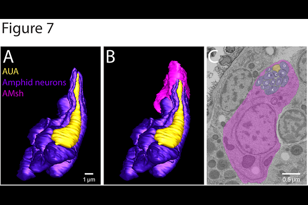
AbstractC. elegans sense organs provide a powerful model for understanding how different cell types interact to assemble a functional organ. Each sense organ is composed of two glial cells, called the sheath and socket, and one or more neurons. A major challenge in studying their development has been the lack of methods to directly observe these structures in the embryo. Here, we mine a recently published high-resolution ultrastructural dataset of a comma-stage embryo that provides an untapped resource for visualizing early developmental events. From this dataset we reconstructed all head sense organs (two amphid (AM), four cephalic (CEP), six inner labial (IL), four outer labial quadrant (OLQ), and two outer labial lateral (OLL)). Symmetric sense organs were at different stages of morphogenesis, allowing us to infer developmental steps by which they form. First, we found that the sheath glial cell begins wrapping its partner neurons at the distal tip of the dendrites where it self-fuses into a seamless tube and then "zippers" down the dendrite. In many cases, sheath glia wrap the progenitors of partner neurons prior to their terminal division. After sheath wrapping has begun, the socket glia wraps the sheath glia circumferentially before presumably elongating to form the mature sheath-socket channel. We also observed transient interactions not found in the mature animal, such as amphid sheath glia wrapping the AUA neuron, that may reflect ancestral relationships. This study demonstrates the value of large public EM datasets that can be mined for new insights, and sheds light on how neurons and glia undergo coordinated morphogenesis.