Seizure Circuit Activity in the Theilers Murine Encephalomyelitis Virus Model of Infection-induced Epilepsy Using Transient Recombination in Active Populations.
Seizure Circuit Activity in the Theilers Murine Encephalomyelitis Virus Model of Infection-induced Epilepsy Using Transient Recombination in Active Populations.
Petrucci, A. N.; Abel, T. N.; Wilcox, K. S.
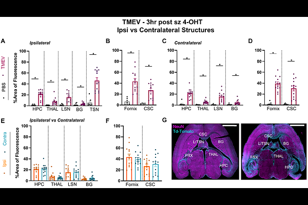
AbstractEpilepsy affects one in twenty-six individuals. A major cause of epilepsy worldwide is viral encephalitis. Central nervous system infections can provoke seizures in the short term and increase the risk of spontaneous, recurrent seizures post-infection. However, the neural mechanisms underlying seizures during acute infection are unknown. These neuronal changes can be studied in C57BL6/J mice infected with Theilers murine encephalomyelitis virus (TMEV). TMEV-infected mice experience seizures 3-8 days post-injection (DPI), clear the virus by DPI 14, and may develop chronic, acquired temporal lobe epilepsy. TMEV may incite seizures during the acute infection period through inflammation, reactive gliosis, and cell death in hippocampal area CA1. Here, we explore the neuronal circuits underlying acute seizures in TMEV-injected mice using c-Fos driven TRAP (targeted recombination in active populations). TRAP mice (c-Fos-CreERT2 x CAG-tdTomato) were injected with PBS or TMEV and gently handled on DPI 5 to induce seizures. 4-OHT was administered to mice either 1.5 or 3 hr after seizures to tag the active cells expressing c-Fos with tdTomato. After 1 week, the mice were sacrificed and whole mouse brains were sectioned and immunostained for tdTomato expression. Percent area of fluorescence was quantified, and comparisons were made between TMEV-injected mice and PBS controls, sites ipsilateral vs contralateral to TMEV injection site, and between sexes. TdTomato expression was elevated in the TMEV-injected mice in the ipsilateral and contralateral hippocampus, thalamus, lateral septal nucleus, basal ganglia, triangular septal nucleus, fornix, and corpus callosum. Critically, the expression pattern suggests that seizures induced on DPI 5 arise from the hilus, dentate gyrus, and CA3 hippocampal subregions. Generalized seizures during acute TMEV infection may have propagated to the contralateral hemisphere via CA3 and the hippocampal commissure. TRAP has not been previously utilized in the TMEV mouse model and these experiments address crucial questions regarding seizure spread during TMEV infection.