Ultra-high field fMRI reveals functional patterns consistent with columnar organisation in human somatosensory cortex
Ultra-high field fMRI reveals functional patterns consistent with columnar organisation in human somatosensory cortex
Dempsey-Jones, H.; York, A.; Shaw, T. B.; Bollmann, S.; Barth, M.; Cunnington, R.; Puckett, A.
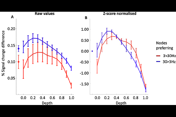
AbstractIn animal models, the primary somatosensory cortex (S1) exhibits columnar organisation, where vertically arranged neurons share functional properties. In humans, however, the thinness and folding of S1 have limited non-invasive investigations of such columnar structures. In this study, we aimed to identify columns in human S1 by delivering alternating bursts of 3 Hz and 30 Hz fingertip vibration while acquiring functional MRI time series at 7 Tesla. Using cortical surface modelling, we identified functional patterns in S1 that showed higher reliability, stronger differential responses, and greater statistical sensitivity than those observed in a frontal cortex control region (p = .001-.012 for reliability; p < .001 for differential signal; p = .004-.011 for sensitivity). Laminar analyses revealed depth-consistent frequency preferences in approximately 20-45% of S1 nodes, a pattern compatible with vertically organised functional structure. Although the relative signal difference between 3 Hz and 30 Hz was small (0.14% signal change), frequency tuning was reliably observed. Taken together, these findings reveal functional patterns in human S1 consistent with aspects of columnar-like organisation, providing non-invasive evidence of fine-scale functional architecture.