Characterising retinal function with optomotor visual performance in P23H rodent models of retinitis pigmentosa
Characterising retinal function with optomotor visual performance in P23H rodent models of retinitis pigmentosa
Brunet, A. A.; Urrutia Cabrera, D.; Wang, L.; Huppert, G.; Chu, S.; James, R.; Harvey, A. R.; Wong, R. C. B.; Carvalho, L. S.
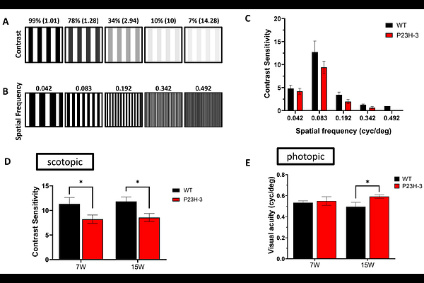
AbstractRhodopsin (RHO) P23H is one of the most common mutations causing autosomal dominant retinitis pigmentosa (adRP), yet the relationship between retinal electrophysiology, structure and visually guided behaviour in rodent models remains unclear. We characterised changes in heterozygous P23H (Sakami line) mice and P23H line 3 (P23H-3) rats using full-field electroretinography (ERG), optomotor response (OMR) assays and, in rats, optical coherence tomography (OCT). ERG assessed rod- and cone-mediated responses relative to wild-type controls, whereas OMR under scotopic and photopic conditions quantified contrast sensitivity and visual acuity. In P23H mice, scotopic ERG responses were significantly reduced from postnatal day 16 and declined further from 4 months. Scotopic OMR contrast sensitivity remained largely preserved until 2 months, and photopic acuity was comparable to wild-type up to 6 months. In 13-week-old P23H-3 rats, ERG amplitudes were significantly reduced, and OCT revealed retinal thinning. OMR showed a decline in contrast sensitivity at 7 and 15 weeks, whereas photopic acuity was maintained. Thus, in both models, electrophysiological and structural abnormalities precede detectable OMR deficits, with implications for the selection of outcome measures in preclinical studies.