Abrupt Pericyte Loss Precedes Endothelial Activation in Cerebral Small Vessel Disease
Abrupt Pericyte Loss Precedes Endothelial Activation in Cerebral Small Vessel Disease
Chagnot, A.; Jaime Garcia, D.; McQuaid, C.; Cholewa-Waclaw, J.; McDade, K.; Dando, O.; Wardlaw, J. M.; Smith, C.; Montagne, A.
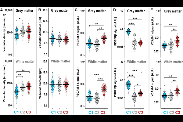
AbstractCerebral small vessel disease (cSVD) is a leading cause of stroke and vascular cognitive impairment, yet the cellular mechanisms underlying microvascular dysfunction in human disease remain incompletely understood. In particular, the relationship between pericyte alterations and endothelial activation, two key features of blood-brain barrier dysfunction, remains unresolved. Here, we performed a quantitative single-vessel analysis of the human cortical microvasculature across increasing cSVD severity and ageing. Using multiplex immunohistochemistry combined with spectral unmixing and automated image analysis, we analysed 11,409 microvascular fragments from post-mortem brain tissue derived from 20 cases. Endothelial cells, pericytes, and endothelial activation were assessed using PECAM-1, PDGFR{beta}, and VCAM-1, respectively. Microvascular density and diameter differed between cortical grey matter and the underlying white matter, with white matter vessels being less dense and wider in controls. While vessel diameter remained stable across disease stages, microvascular density increased with cSVD severity and age in the white matter. At the molecular level, PDGFR{beta} signal decreased markedly with increasing cSVD severity, consistent with progressive pericyte loss. This reduction was observed in both grey and white matter and correlated with disease severity and age. Notably, intermediate disease groups displayed marked heterogeneity, with vessels exhibiting either preserved or near-complete pericyte coverage, suggesting a potentially bimodal transition. In parallel, endothelial markers PECAM-1 and VCAM-1 increased significantly with disease severity, reflecting endothelial activation. Unsupervised Gaussian mixture clustering of marker expression identified three vascular states characterised by (i) preserved pericytes with low endothelial activation, (ii) marked pericyte loss without endothelial activation, and (iii) combined pericyte depletion and endothelial activation. These clusters broadly aligned with clinical severity but revealed intermediate states not captured by post-mortem diagnosis alone. Together, these findings suggest that pericyte loss and endothelial activation are partially dissociated processes that occur in a sequential progression in human cSVD, supporting pericyte dysfunction as an early event and highlighting it as a potential therapeutic target in microvascular disease.