Electron tomography reveals mitochondrial network and cristae remodelling during cell differentiation in the human placenta
Electron tomography reveals mitochondrial network and cristae remodelling during cell differentiation in the human placenta
Acharya, S.; Hanssen, E.; Botha, V. B.; Smith, T. M.; Jayatissa, S.; Trifunovic, Z.; Bartho, L. A.; Schjenken, J. E.; Kaitu'u-Lino, T. J.; Perkins, A. V.; James, J. L.; Pringle, K. G.; Bouwer, J. C.; Smith, R.; Fisher, J. J.
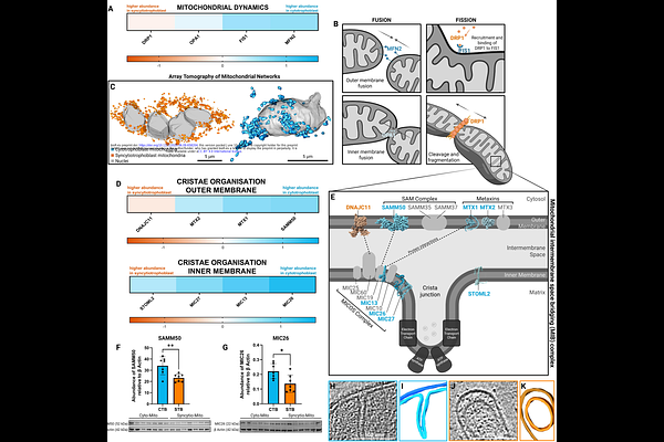
AbstractMitochondria adapt their structure through fusion and fission, yet how their morphology and cristae architecture alter as cells differentiate remains unclear. The human placenta is an ideal model for studying mitochondrial diversity within a single tissue. As epithelial trophoblast cells of the placenta differentiate, their accompanying mitochondria morphologically and functionally transform. We used array tomography to characterise mitochondrial volume and network complexity. Cryo-electron tomography revealed two distinct subpopulations of mitochondria within progenitor cytotrophoblasts, and a singular, homogeneous population in the differentiated syncytiotrophoblast. We showcased the 3D topology of individual cristae through a standardised metric of \'\'curvedness\'\'. Proteomic analysis of isolated mitochondria identified reduced dynamics proteins and cristae organisation complexes, accompanied by subunit variations within the electron transport chain, ATP synthase, and altered supercomplex abundance. This study highlights the advantages of combining multimodal imaging with mechanistic insights, to elucidate the process of mitochondrial morphology and cristae architecture remodelling as cells differentiate.