Development of noninvasive imaging to measure spontaneous pain in mice
Development of noninvasive imaging to measure spontaneous pain in mice
Yuan, S.; Liew, J. Y.; Pal, A.; Wang, J.; Green, D. P.; Motamedi, M.; Tang, S.-J.
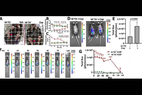
AbstractObjectively measuring pain in laboratory animals is essential for pain research and analgesic development. Despite the development of various behavioral tests to measure evoked pain in animal models, measuring spontaneous pain remains challenging. To address this unmet need, we developed a novel imaging approach to detect spontaneous nociception in animal pain models. To do this, we generated a Bacterial Artificial Chromosomes transgenic mouse that expresses Redquorin under the murine synapsin 1 promoter. Redquorin is a fusion protein consisting of chimera and a 2x tandem dimer Tomato Aequorin (tdTA), which emits long wavelength bioluminescence from activated neurons in the presence of coelenterazine. This luminescence can penetrate tissues and form a projected image on the body surface that can be detected with a spectrum In Vivo Imaging System, thus creating a Nociceptive Neuronal Activity Imaging mouse. We used the tdTA mice to image bioluminescence in the spinal regions as a surrogate of spontaneous pain induced by capsaicin, the HIV-1 envelope glycoprotein gp120, and spinal nerve ligation. Results show that Redquorin-emitted bioluminescence is a sensitive optical surrogate to measure spontaneous pain. This approach offers a new method to measure spontaneous pain in animal models for basic and translational research.