Apical3DTip: Elliptic Cross-section-based Reconstruction for the Embryo Initial Cell of Arabidopsis

Apical3DTip: Elliptic Cross-section-based Reconstruction for the Embryo Initial Cell of Arabidopsis
Nonoyama, T.; Kang, Z.; Hanaki, Y.; Itagaki, Y.; Matsumoto, H.; Kimata, Y.; Tsugawa, S.; Ueda, M.
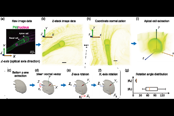
AbstractBackgroundCell geometry plays a central role in determining division orientation and body axis formation during early embryogenesis in Arabidopsis thaliana. However, quantitative analysis of dynamic three-dimensional (3D) morphology remains challenging because live-imaging studies often rely on two-dimensional (2D) projections, while existing 3D reconstruction approaches, including mesh-based methods, often lose the original orientation information relative to the ovule and require labor-intensive mesh correction. In addition, embryo positional fluctuation caused by floating in liquid medium and continuous growth makes it difficult to analyze temporal morphological changes within a common coordinate system. ResultsWe developed a robust framework for quantitative 3D and four-dimensional (4D; 3D + time) analysis of embryo initial cell (apical cell) morphology. The method first establishes a standardized 3D coordinate system by normalizing cell orientation based on the bottom plane and the optical axis of the observation. Cell morphology is then reconstructed through ellipse-based approximation of serial cross-sections extracted from stacked imaging data, enabling accurate geometric characterization without the need for complex surface mesh reconstruction. To evaluate shape anisotropy, we quantified the apical cell shape in 3D. The framework further supports the characterization of volumetric features of subsequent division, providing a basis for quantifying 3D embryogenesis. ConclusionOur framework provides a simple and noise-reduced approach for quantitative analysis of living cell morphology in 3D. We named the integrated method of combining coordinate normalization with elliptical cross-section-based reconstruction Apical3DTip. This method enables consistent comparison of cell shapes without extensive manual corrections. The method overcomes key limitations of 2D projection-based and mesh-dependent analyses and offers a practical platform for quantifying cell shape and daughter cell shapes in 3D. More broadly, it provides a quantitative foundation for exploring the relationship between cell geometry, morphodynamics, and developmental patterning in living plant embryos.